ModuleSci, South Korea
Normal W-SEM
Model: PicoEye-100 (PE-100)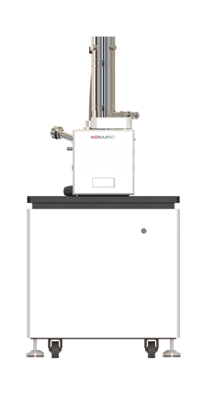
Key Features
- Compact tungsten filament Scanning Electron Microscope built for ease of operation, low cost of ownership and broad applicability
- Suited to fracture analysis of metallic materials, phase-separation observation in polymers, and failure root-cause tracing
- Resolution of 3 nm at 30 kV, with simultaneous high-quality SE and BSE imaging plus image mixing
- Optical navigation and sample-chamber monitoring for rapid sample positioning
- One-click automatic adjustment and fast vacuum pumping simplify daily inspection
- Low vacuum mode observes non-conductive materials directly, without conductive coating
- Supports multiple detector options and image-stitching module expansion
Specifications
| Parameter | Specification |
|---|---|
| Emission source | Pre-aligned tungsten filament electron gun |
| Resolution | 3.0 nm |
| Magnification (photo) | 30× ~ 300,000× |
| Accelerating voltage | 1~30 kV |
| Detector | Secondary Electron Detector (ETD); optional Backscattered Electron Detector (BSED), EDS |
| Image format | TIFF, JPG, BMP, PNG |
| High vacuum mode | ≤5×10⁻⁴ Pa |
| Low vacuum mode (optional) | 6~150 Pa |
| Automatic stage | 5-axis |
| Stage moving range | X: 0~50 mm, Y: 0~50 mm, Z: 3~50 mm |
| Main unit dimensions | 670(W) × 780(D) × 1450(H) mm |
| Weight | 310 kg |
| Room requirements | 2m × 2.5m × 2m |
| Power supply | AC 110/220V (±10%), 50/60Hz, 1.5kW |
| Temperature & humidity | 20±5°C, ≤60%RH |
A compact tabletop sibling (PV-100P) and a field-emission model (PE-300) are also available in the ModuleSci SEM range.
Application Examples
A sample of imaging results produced on the PE-100.
Ceramic
5kV, SE imaging
Steel Inclusions
15kV, SE imaging
Drosophila Compound Eye
15kV, SE imaging
Tin Ball
20kV, SE imaging
Semiconductor Chip
20kV, BSE imaging
Mask
30kV, SE imaging
Intelligent Operating Software
ModuleSci’s self-developed SEM software captures an image in three guided steps: mount and set the sample, take a navigation image while the chamber evacuates, then navigate to the region of interest and capture the SEM image. Automatic brightness/contrast, focus and astigmatism adjustment run in the background, with live chamber monitoring throughout.
Photo-assist tools include dimension measurement, image mixing and gesture-based quick navigation, with optional image stitching for wide-field views.
Software at a Glance
- Workflow
- Mount sample → navigate & evacuate → locate ROI & capture
- Languages
- Korean, English, Chinese, French, German, Russian
- Photo-assist tools
- Dimension measurement, image mixing, gesture navigation, optional image stitching
Get A Free Instrument Consultation!
We can suggest products to our customers to make them the leaders in their respective industries.